

Issue No. 151
Summarized from Journal of Clinical Periodontology, Volume 53, Issue 7, July 2026, 1005-1015
Editor: James Deschner, chair, EFP scientific affairs committee
Can orthodontic tooth movement start one week after regenerative surgery?
Authors: Pal Nagy, Peter Windisch, Balint Nemes, Peter Schupbach, Andrea Dobos, Andreas Stavropoulos
Background
Interdisciplinary treatment combining periodontal regeneration and orthodontic tooth movement (OTM) is increasingly used to treat pathologic tooth migration associated with periodontitis. Preclinical data suggest that orthodontic movement into regenerated sites can be performed without jeopardizing the formation and the gain in new periodontal attachment apparatus and may even enhance bone apposition through tensile-site bone apposition.
However, human histological evidence is scarce. It remains unclear how non-resorbable or slowly resorbable bone substitutes behave when subjected to early orthodontic movement, and whether these orthodontic forces alter the ratio of new bone to residual graft material in humans.
Aims
The aim was to histologically and histomorphometrically evaluate the first 20 consecutive cases from a randomized controlled clinical trial that investigated the effects of early OTM on the healing of periodontal intrabony defects treated with guided tissue regeneration (GTR) using deproteinized bovine bone mineral (DBBM).
Materials and methods
- Following step 1 and step 2 periodontal therapy, the first 20 patients from the randomized controlled trial were selected for histological analysis.
- Participants: Adults aged 18–65 with pathologic tooth migration requiring both regenerative and orthodontic therapy. Intrabony defects with the following morphology:
- intrabony depth (≥4mm)
- Wide defects (radiologic angulation >25°)
- Non-contained defects (one or two bony walls)
- Regenerative periodontal surgeries were performed by a sole certified periodontist at a single university centre. GTR with a coronally advanced flap design was performed. The intrabony defects were filled with DBBM and covered with a resorbable collagen membrane. A premade stent was fitted with a trephine and calibrated for re-entry at nine months.
- After surgery, the sole operator generated a random allocation sequence and assigned the patients to two groups:
- Test group (n=11): early OTM (seven days post-op) was performed on the study tooth by an orthodontist. Subjects were divided into sub-groups:
- T1 (n=6): OTM directed away from the previous defect
- T2 (n=5): OTM towards the previous defect
- Test group (n=11): early OTM (seven days post-op) was performed on the study tooth by an orthodontist. Subjects were divided into sub-groups:
- Control group (n=9): received no orthodontic treatment, study tooth remained splinted during the healing period of nine months.
- All patients were given monthly supportive care until surgical re-entry at nine months following surgery by the same surgeon.
- At re-entry, the previously fitted stent and trephine (2mm diameter) were utilized to harvest a sample as close to the root surface as possible, while avoiding damage to the regenerated site. The depth of the sample was determined by calculating the amount of bony fill of the intrabony defect.
- The histologist was blinded to the study protocol and group allocation.
- Main outcomes included histological and histomorphometric analyses of the bone sample.
- Secondary outcomes included clinical and radiological parameters.
Results
- From the 20 collected samples, 19 biopsies were successfully evaluated (one test sample was damaged). Four patients (three controls, one test) experienced early wound dehiscence, which resolved with secondary healing.
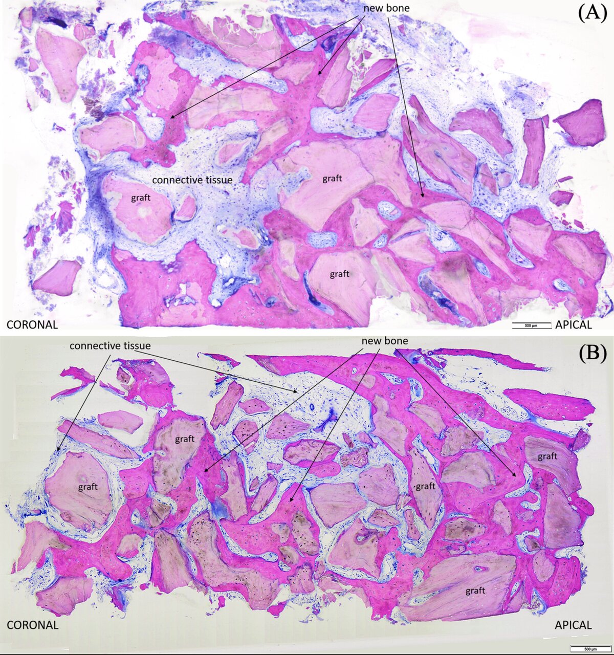
- New bone formation: The test group (OTM) showed significantly higher new bone formation compared to the control group (33.9% ± 13.0% vs. 17.4% ± 9.2%, p=0.011).
- Residual graft: The test group exhibited a significantly lower ratio of residual DBBM graft percentage than the control group (16.3% ± 9.2% vs. 33.2% ± 3.7%, p=0.001).
- Soft tissue: Connective-tissue components and bone-marrow percentage ratio did not show significant differences between groups (49.7% vs. 49.4%, p=0.74).
- Histological features:
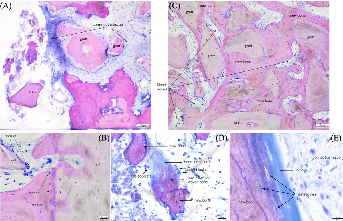
- Control samples: DBBM served as a scaffold, with new bone largely confined to the apical and middle thirds, while coronal graft particles were embedded in connective tissue. The new bone interlinks the xenograft particles with osteoblast cell activity, osteoid production, and the formation of woven-type bone.
- Test samples: In both T1 and T2, similar osteoclastic activity, osteoid and woven bone formation were also observed. Cutting cones (osteoclastic cell functions create resorption lacunae inside the graft materials) were observed. In some T2 samples, complete disappearance of the graft particles, bone in-growth into the graft particles, and encapsulation of the graft particles by the connective tissues were also found.
Limitations
- The bone samples from the trephine drills captured only the alveolar bone adjacent to the tooth to avoid damage to the regenerated site without sampling tissues from the periodontal attachment apparatus, because of ethical concerns. This may preclude any conclusions about true periodontal regeneration (formation of new cementum and periodontal ligament).
- The main findings on histomorphometric analysis relies on a standardized application of the DBBM into the defects in surgery and a standardized method to sample bone at re-entry. Multiple roles were handled by the same sole operator. Potential performance bias remains at the nine-month surgical re-entry, even though the randomization sequence and allocation were performed after the initial surgery.
- The intervention, orthodontic mechanics and force regime—which were routinely planned on a case-by-case basis—is by nature difficult to standardize or replicate.
- The relatively small sample size with unclear study power and lack of repeated measurement values reported for the histomorphometric analysis.
- Limited conclusions regarding the effect of OTM on the long-term clinical and tangible patient outcomes of intrabony defects that received GTR can be drawn solely from the current histological and histomorphometric evaluation.
Conclusions and impact
- Early OTM about a week after regenerative surgery with native resorbable collagen membrane and DBBM in intrabony defect does not appear to jeopardise periodontal healing at nine months.
- Orthodontic forces applied early in the healing phase appear to promote bone remodelling, resulting in relatively more new bone formation compared to DBBM, while the total amount of bone remained similar.
- From the histological standpoint, for patients needing both regeneration and orthodontics, a long healing period before starting tooth movement may not be necessary for patients receiving GTR. This may reduce the treatment timeline in periodontal-orthodontic cases.
Until more long-term studies confirm this, the recommendation of the EFP clinical-practice guideline on the treatment of stage 4 periodontitis of four to six weeks remains reasonable.
Rapporteurs: Ja Young Jane Lee, Yi Ning Low, Chin Hung Lai, and Katherine Lau supervised by Professor Chris T. C. Fok and Professor George Pelekos
Affiliation: Postgraduate programme in periodontology, University of Hong Kong
With kind permission from Wiley Online Library. Copyright © 1999-2026 John Wiley & Sons, Inc. All rights reserved
EFP partners









Sign up to our newsletter
The EFP publishes a free monthly email newsletter with the latest news about the federation's activities, its publications, and its campaigns.
