

Issue No. 155
Summarized from Journal of Clinical Periodontology, Volume 53, Issue 11, November 2026, 1691-1701
Editor: James Deschner, chair, EFP scientific affairs committee
How do keratinized mucosa and mucosal thickness affect long-term implant outcomes?
Authors: Emilio Couso-Queiruga, Clemens Raabe, Andrea Roccuzzo, Giovanni E. Salvi, Manrique Fonseca, Vivianne Chappuis
Read the full JCP paper
Background
The peri-implant soft-tissue phenotype, primarily characterized by keratinized mucosa width (KMW) and mucosal thickness (MT), represents an important factor that potentially influences peri-implant health, tissue stability, and aesthetic outcomes.
Inadequate KMW has been associated with increased plaque accumulation, bleeding on probing (BOP), peri-implant soft-tissue dehiscence (PSTD), brushing discomfort, and crestal bone loss (CBL). These associations appear particularly relevant in patients with irregular adherence to supportive peri-implant care.
Similarly, reduced MT has been associated with a higher prevalence of soft-tissue dehiscence, whereas thicker mucosa and soft-tissue augmentation procedures may contribute to improved clinical and radiographic stability.
Nevertheless, evidence regarding the association between KMW, MT, and peri-implant diseases remains inconsistent. While some studies suggest a protective effect of adequate soft-tissue dimensions, others have failed to demonstrate significant associations. Furthermore, the long-term influence of these parameters in patients who maintain adequate oral hygiene and receive regular supportive peri-implant care remains insufficiently investigated.
Aims
The primary aim of this 25-year cohort study was to investigate the association between KMW and CBL. Secondary aims were to assess the associations between both KMW and MT and peri-implant clinical parameters, tissue conditions, and patient-reported discomfort during brushing. Longitudinal assessment was applied to KMW, while MT was measured only at the 25-year follow-up.
Materials and methods
Study design: This cohort study was conducted at the Department of Oral Surgery and Stomatology of University of Bern (Switzerland). Adult patients from a previously reported implant cohort (Buser et al., 2012) were invited for re-examination. The 10-year data were analysed retrospectively for the present study, whereas only the 25-year examination was prospectively planned and conducted under the current study protocol.
Eligibility criteria: Only implant-supported prostheses (ISPs) with available data at both the 10- and 25-year follow-ups were included.
Data collection:
- Clinical parameters were recorded at implant sites at 10- and 25-year examinations: probing depth (PD), BOP, suppuration on probing (SOP), plaque index, and KMW (measured at the mid-facial aspect of each implant).
- MT was assessed only at 25 years: 3mm apical to the mucosal margin using a transgingival probing method.
- Standardized digital periapical radiographs were taken to assess CBL and implant-supported prosthesis fit, while standardized intraoral photographs were captured to evaluate the presence of a PSTD.
- Peri-implant health, peri-implant mucositis, and peri-implantitis were defined based on the 2018 Classification of Periodontal and Peri-implant Diseases and Conditions and the EFP’s clinical-practice guidelines.
- Patient-reported brushing discomfort was assessed before the 25-year examination and classified dichotomously as “yes” or “no”.
Statistical analysis: Demographic, implant- and prosthetic-related characteristics, and study outcomes were stratified by the three KMW groups (0mm, < 2mm, and ≥ 2mm). Associations of MT stratified as thin (< 2mm) and thick (≥ 2mm) and KMW with continuous outcomes were examined using robust linear mixed-effects models. Binary outcomes were analysed using generalized estimating equation models, and nominal categorical outcomes were assessed using multinomial-Poisson generalized linear mixed models.
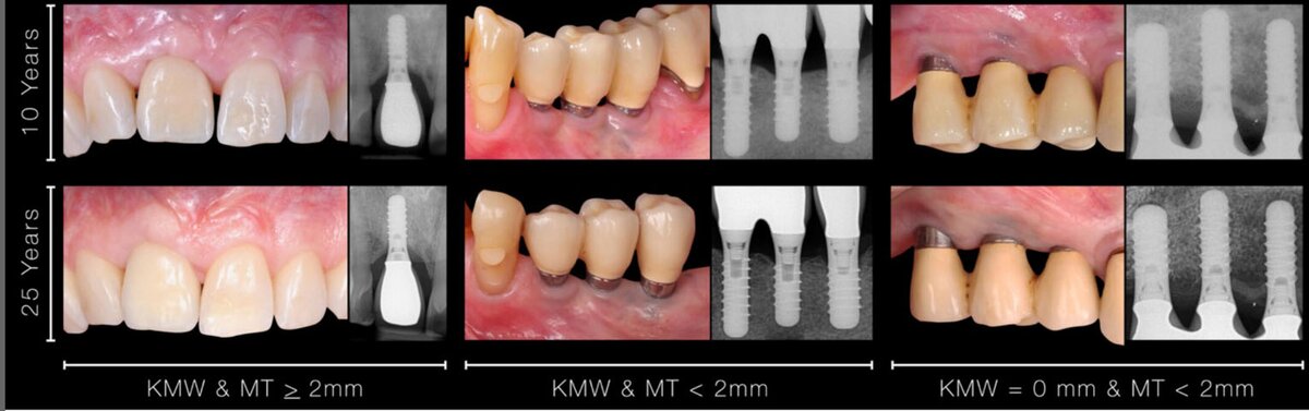
Results
Study population: 149 patients and 235 implant-supported prostheses were available for analysis at both 10 and 25 years.
Keratinized mucosal width:
- Crestal bone loss: At 10 and 25 years, statistically significantly greater CBL was observed in ISPs with 0mm KMW (0.29 ± 0.79mm and 0.62 ± 1.04mm) compared with ISPs presenting < 2mm (−0.18 ± 0.80mm and 0.01 ± 0.77mm) and ≥ 2mm KMW (−0.18 ± 0.81mm and 0.24 ± 1.2mm).
- Peri-implant diseases: At the 25-year follow-up, the prevalence of peri-implantitis was four cases (11.1%) in the 0mm group, zero cases in the < 2mm group, and 12 cases (7.1%) in the ≥ 2mm group. Peri-implant mucositis was the predominant diagnosis, affecting 29, 27, and 128 ISPs in the 0mm, < 2mm and ≥ 2mm groups, respectively. Differences between KMW groups were not statistically significant for either condition (p > 0.05).
- Peri-implant soft-tissue dehiscence: At both time points, the 0mm KMW group exhibited a statistically significantly higher prevalence of PSTD (97.2%) (p < 0.05) and similarly, the visibility of the implant platform was most frequent in the 0mm group.
Mucosal thickness:
- Crestal bone loss: Although a lower mean CBL was observed at both time points for thicker MT group, the differences were not statistically significant between thin and thick mucosa phenotypes.
- Peri-implant diseases and clinical parameters: MT was not statistically associated with peri-implant mucositis or peri-implantitis at either time point. Thick (≥ 2 mm) MT was not statistically associated with lower mean PD, BOP, and SOP values; contrarily, the thicker MT group was associated with higher plaque accumulation at 25 years (p = 0.01).
- Peri-implant soft-tissue dehiscence: The thicker MT group was associated with lower risk of PSTD (at 10 and 25 years) and visibility of implant platform (at 25 years only).
Brushing discomfort: Discomfort was observed in 11.4% of ISPs in the 0mm KMW group, 3.4% of ISPs in the < 2mm group and 2.4% in the ≥ 2mm group, without significant differences between KMW or MT groups.
Limitations
- Since it is a cohort study, the reported associations between KMW, MT, and implant-related outcomes may be influenced by residual confounding factors during the long follow-up period.
- Only tissue-level titanium implants were analysed, and the cohort consisted of a Swiss population enrolled in regular supportive peri-implant care with generally adequate oral hygiene. Therefore, the findings may not be fully generalizable to other implant systems or to patients with less stringent maintenance protocols and oral-hygiene habits.
- Implant failures were not considered.
- MT was assessed only at the 25-year follow-up, precluding the evaluation of temporal associations.
- The categorization of KMW (0mm, < 2mm, ≥ 2mm) and MT (< 2mm vs. ≥2 mm), and the dichotomous assessment of brushing discomfort may have led to loss of information.
- MT was measured at a single apico-coronal level at the facial site using a transgingival probing method, which may not fully represent the three-dimensional soft-tissue phenotype.
Conclusions and impact
- Absence of KMW was associated with higher crestal bone loss as well as an increased risk of peri-implant soft-tissue dehiscence and visibility of implant platform.
- Thick (≥ 2mm) MT was associated with lower risk of peri-implant soft-tissue dehiscence and visibility of implant platform.
- Brushing discomfort was not statistically associated with KMW and MT.
- Peri-implant health, peri-implant mucositis ,and peri-implantitis were not significantly related to KMW and MT.
These findings highlight the clinical relevance of the peri-implant soft-tissue phenotype, suggesting that the presence of keratinized mucosa and thick mucosa may be associated with long-term peri-implant tissue stability, particularly a lower risk of soft-tissue dehiscence and, for keratinized mucosa only, less crestal bone loss. In well-maintained patients, neither was significantly associated with peri-implant disease or brushing discomfort.
Rapporteurs: Camilla Cavalcanti and Gessica Pettinari, supervised by Professor Francesco Cairo
Affiliation: Postgraduate programme in periodontology, University of Florence, Italy
With kind permission from Wiley Online Library. Copyright © 1999-2026 John Wiley & Sons, Inc. All rights reserved
EFP partners








Sign up to our newsletter
The EFP publishes a free monthly email newsletter with the latest news about the federation's activities, its publications, and its campaigns.
