

Issue No. 154
Summarized from Journal of Clinical Periodontology, Volume 53, Issue 10, October 2026, 1565-1574
Editor: James Deschner, chair, EFP scientific affairs committee
How do platelet concentrates and collagen substitutes affect palatal wound healing?
Authors: Stefan P. Bienz, Anne C. Fischer, Leonardo Mancini, Franz J. Strauss, Raphael M. Ferrari, Sonja Hitz, Ronald E. Jung, Daniel S. Thoma.
Background
Mucogingival surgery is employed to improve the quality and/or quantity of periodontal and peri-implant soft tissues, promoting tissue homeostasis and maximizing aesthetic outcomes. Nevertheless, mucogingival surgery is often associated with postoperative discomfort and potential complications that affect healing at the donor and/or the recipient site, necessitating the use of adjunctive agents that could enhance tissue repair and patient comfort.
Several adjunctive approaches have been proposed, including collagen-based wound dressings and autologous platelet concentrates, such as platelet-rich fibrin (PRF) and plasma rich in growth factors (PRGF). These blood-derived products have been shown to modulate tissue inflammation while promoting biological mechanisms of wound healing at the cellular and molecular level.
In contrast, collagen-based substitutes support the wound healing process by providing the mechanical stability of the blood clot and the reconstructed tissues. While studies have shown that platelet concentrates remain promising for their biological effects in soft-tissue healing, there is inconsistent evidence for their clinical effectiveness, especially when compared with unassisted healing.
As patient-reported outcomes have become increasingly important in periodontal and implant therapy, platelet concentrates have attracted interest for their potential to reduce postoperative pain. Therefore, assessing wound closure and postoperative discomfort is important to determine whether these adjunctive strategies provide benefits over spontaneous healing.
Aims
This study aimed to determine the effectiveness of three experimental strategies—platelet-rich fibrin (PRF), plasma rich in growth factors (PRGF), and a collagen-based substitute— compared with spontaneous healing in standardized palatal wounds. Wound-closure progression and patient-reported postoperative pain were evaluated during the first 14 days of healing.
Materials and methods
- Study design: This randomized controlled clinical trial employed a within-subject experimental design of 20 systemically healthy participants. Each participant received all four treatment modalities, randomly allocated to separate palatal sites.
- Eligibility criteria: Participants were aged 18–80, with good oral hygiene (plaque index <30%), free of periodontitis (probing depths ≤3 mm), and suitable anatomy of the palate to facilitate the creation of standardized wound defects.
- Surgical procedure: A biopsy punch was used to create four circular palatal defects (6mm in diameter and 3mm in depth) per participant, with two defects on each side of the palate having a minimum of 3mm distance in-between the adjacent wounds and teeth.
- Treatment groups were as follows:
- PRF: Autologous platelet-rich fibrin membrane prepared without anticoagulants and precut to fit the defect.
- PRGF: Autologous plasma-rich in growth factors clot prepared using sodium citrate as an anticoagulant and precut to fit the defect.
- CS: Collagen-based substitute membrane (Geistlich Mucograft), trimmed to fit the defect.
- Control: Unassisted healing without additional wound dressing.
- Wound stabilization: All sites received crisscross sutures, including the control sites, to stabilize the materials, where applicable, and the blood clot.
- Postoperative care: Participants gently rinsed with 0.2% chlorhexidine mouthwash twice daily until suture removal on the fifth day. Ibuprofen (600 mg) was prescribed as needed.
- Outcome assessment: Standardized photographs were obtained at baseline and at two, five, seven, nine, 12, and 14 days postoperatively. The primary outcome was the percentage of remaining open-wound area, quantified using image-analysis software. Pain was assessed separately for each site using an 11-point numerical rating scale (0–10).
- Statistical analysis: Longitudinal statistical models evaluated treatment-related differences, while accounting for repeated measurements within participants.
Results
- Study population and adverse events: All 20 participants were enrolled. One participant missed two follow-up visits. Four subjects experienced premature suture loss without displacement of wound dressings or clots. No other adverse events were reported.
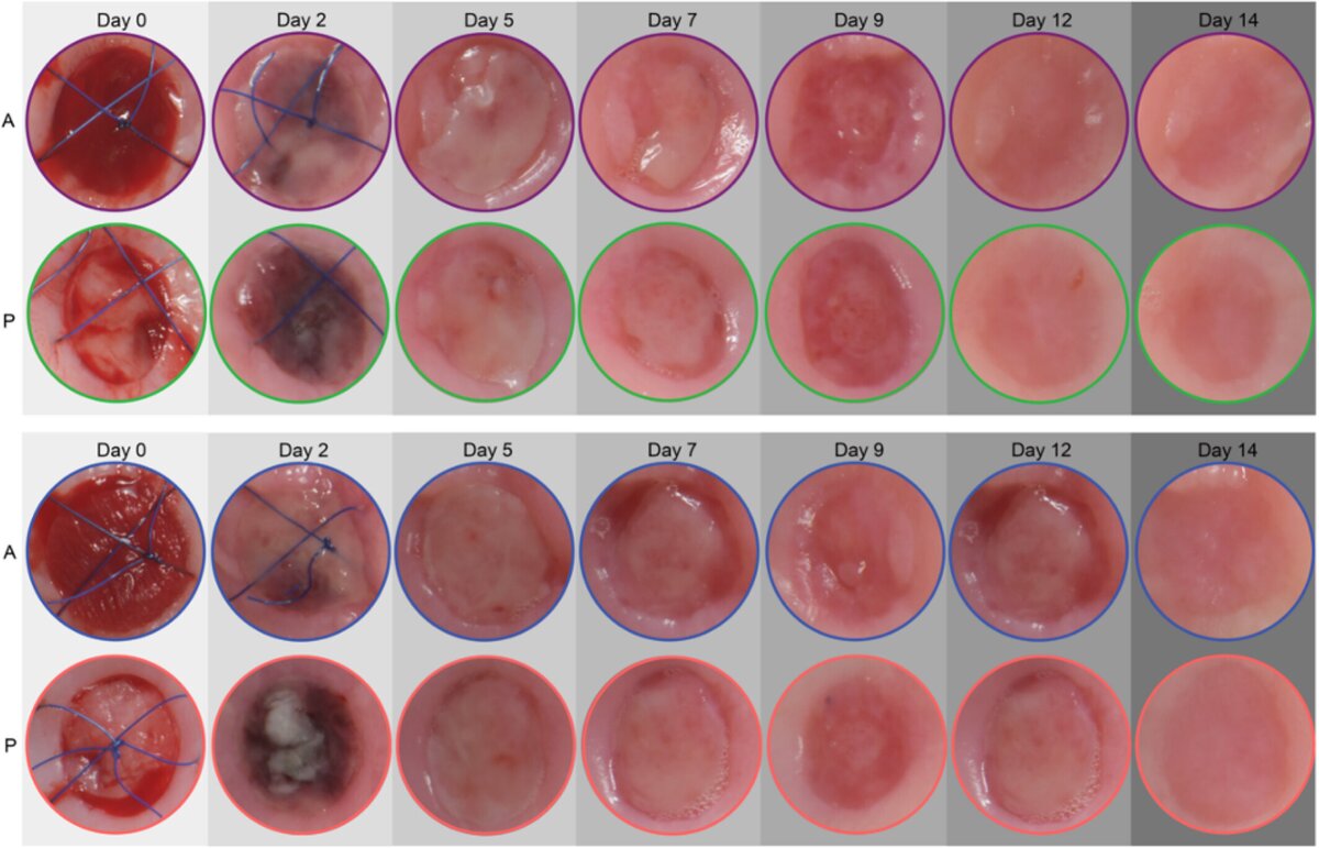
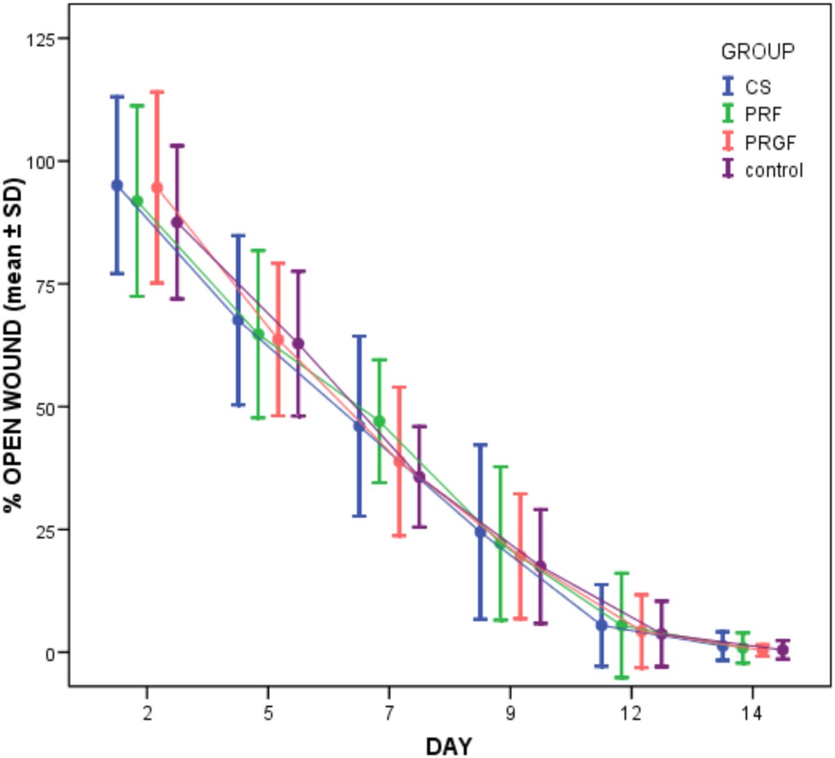
- Wound-healing progression: The remaining open-wound area progressively decreased in all four groups throughout the 14-day observation period. Temporary wound enlargement was observed in some defects on Day 2.
- Day 7 findings: The greatest inter-group differences were noted on Day 7. The remaining open-wound area was 47.0% ± 12.5% for PRF, 38.8% ± 15.1% for PRGF, 46.0% ± 18.3% for CS (collagen substitute), and 35.7% ± 10.2% for spontaneous healing.
- Treatment comparisons: Across visits and anatomical locations of defects, unassisted healing demonstrated significantly smaller remaining wound areas compared with PRF (p = 0.018; Cohen's d = 0.67) and CS (p = 0.042; Cohen's d = 0.60).
- Day 14 findings: Complete wound closure was achieved in 85% of defects. Mean residual wound areas were 0.9% for PRF, 0.4% for PRGF, 1.3% for CS, and 0.5% for controls.
- Anatomical location: Healing progression was significantly influenced by the interaction between time, treatment, and wound location (p < 0.001). Anterior defects showed a tendency for faster healing than posterior defects.
- Postoperative pain: Pain decreased significantly over time (p < 0.001), without significant differences between treatment groups (p = 0.737). Anterior sites tended to be more painful than posterior sites (p = 0.084).
Limitations
- Limited sample size: The inclusion of only 20 participants may have reduced statistical power to detect smaller but potentially meaningful differences between treatments.
- Limited generalizability: The predominantly young, systemically healthy population and small standardized palatal defects may not reflect healing outcomes in larger surgical wounds or patients with compromised healing.
- Short follow-up period: The 14-day observation period did not allow assessment of longer-term tissue maturation or healing quality.
- Incomplete blinding: The control sites were identifiable during photographic assessment, introducing potential observer bias.
- Non-standardized analgesic intake: Differences in pain medication consumption may have influenced patient-reported pain scores.
- Potential biological interference: The proximity of the four defects may have allowed biological effects from neighbouring treatment sites to influence healing, potentially affecting comparisons.
- Assessment limitations: Photographic measurements evaluated surface wound closure without providing histological information about tissue regeneration.
Conclusions and Impact
- Unassisted healing demonstrated slightly faster wound closure than the adjunctive treatments, with statistically significant differences observed for PRF and CS but not PRGF.
- The application of PRF, PRGF, and CS did not provide additional benefit for early wound closure, suggesting limited added value in small, standardized palatal defects.
- Postoperative pain decreased progressively across all treatment groups, with no statistically significant differences between the interventions.
- Wound location appeared to influence healing progression and patient discomfort, with anterior palatal sites tending to heal more quickly but with greater pain perception than posterior defects.
- The favorable healing capacity of healthy palatal tissues may reduce the need for additional biomaterials in uncomplicated wounds.
- Further clinical investigations are necessary to establish whether these materials provide greater benefits in larger defects or in patients with compromised healing.
In small-sized, standardized wounds created on the palate of healthy individuals, PRF, PRGF, and CS were not found to be superior to unassisted healing regarding wound closure or postoperative pain. These findings indicate no clinically relevant additional benefit of these adjunctive biomaterials, so that their routine use is therefore not suggested in these clinical scenarios. Further research is needed to establish their potential benefits in more complex cases.
Rapporteurs: Iason Chatzimentor, Amalia Michou, Vasilis Tilaveridis, and Vasiliki Savva, with Danae Apatzidou (programme director)
Affiliation: Postgraduate programme in periodontology, Aristotle University of Thessaloniki, Greece
With kind permission from Wiley Online Library. Copyright © 1999-2026 John Wiley & Sons, Inc. All rights reserved
EFP partners







Sign up to our newsletter
The EFP publishes a free monthly email newsletter with the latest news about the federation's activities, its publications, and its campaigns.
