

Issue No. 152
Summarized from Journal of Clinical Periodontology, Volume 53, Issue 8, August 2026, 1289–1300
Editor: James Deschner, chair, EFP scientific affairs committee
Identifying prognostic factors for tooth survival in endo-perio lesions
Authors: Fumihiko Kimura, Risako Mikami, Koji Mizutani, Daisuke Kido, Kohei Takeda, Keita Nakagawa, Shu Takemura, Hiromi Kominato, Natsumi Saito, Tatsuro Seike, Eri Sakaniwa, Ayu Sugiyama, Tomoki Nogami, Satoshi Watanabe, Jun Aida, Takanori Iwata
Background
Endodontic-periodontal lesions (EPLs) are complex pathologies, characterized by deep periodontal pockets that reach or involve the root apex, that result from pathological communication between the pulp and periodontal tissues. These lesions pose a significant clinical challenge because of their multifactorial nature and the compromised prognosis associated with combined endodontic and periodontal involvement.
The 2017 World Workshop classification of periodontal diseases and conditions introduced a grading system for EPLs based on the number and extent of deep periodontal pockets. However, evidence on long-term tooth survival remains scarce, as most available studies are small-scale and have observation periods of one year or less.
Although radiographic alveolar bone loss and periapical radiolucency have been suggested as potential predictors of treatment outcomes, the prognostic relevance of the overall radiographic pattern of bone loss has not been adequately explored.
A standardized and validated prognostic model for teeth with EPLs is still lacking, and larger studies with longer follow-up periods using survival analysis are needed to better understand which clinical and radiographic factors influence tooth retention after treatment.
Aim
This study sought to identify pretreatment factors that predict tooth survival in teeth affected by EPLs, with particular focus on whether the radiographic shape of bone loss could serve as an additional prognostic indicator alongside established clinical parameters.
Materials & methods
Retrospective cohort study conducted at the Periodontal Clinic of Tokyo Medical and Dental University Hospital (April 2015–March 2021).
Inclusion criteria:
- Age ≥20 years.
- Teeth diagnosed with EPL, presenting with a deep periodontal pocket extending to or approaching the root apex with corresponding radiographic alveolar bone loss.
- Baseline probing depth (PD) ≥5mm.
- Minimum follow-up period of six months after root-canal filling (RCF).
Exclusion criteria:
- Smoking.
- Unavailable baseline periodontal records or radiographs.
- Transplanted teeth.
- Teeth with suspected root damage (cracks, fractures, or external root resorption).
EPL classification: EPLs were classified in patients with or without periodontitis and graded (1–3) based on the number and extent of deep periodontal pockets, according to the 2017 classification.
Radiographic assessment:
- Bone loss was calculated as the mean of mesial and distal measurements and expressed relative to root length.
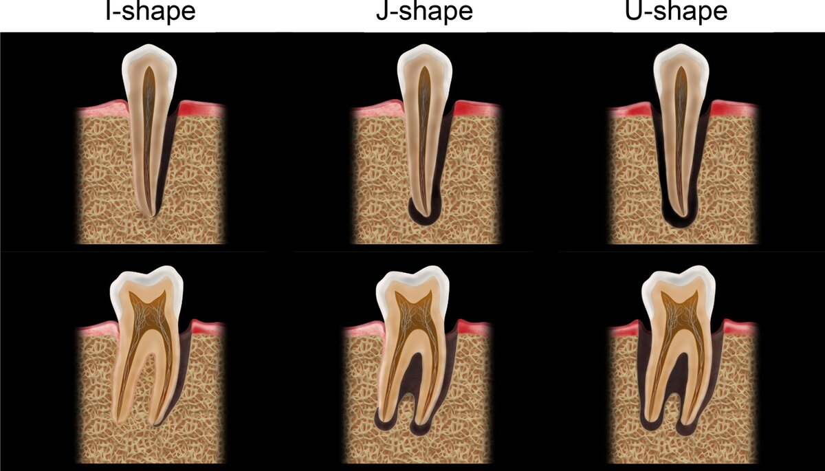
- Periapical radiographs were evaluated by two calibrated periodontists using the Periapical Index (PAI) and the I-J-U subclassification:
- I, unilateral intrabony defect without periapical radiolucency.
- J, unilateral intrabony defect with periapical radiolucency.
- U, bilateral intrabony defects with periapical radiolucency (κ = 0.83).
Baseline clinical parameters: Probing depth, bleeding on probing, tooth mobility, furcation involvement, periodontal diagnosis, and history of endodontic treatment were evaluated.
Treatment:
- Endodontic treatment was performed first.
- Non-surgical periodontal therapy was initiated after about three months.
- Following re-evaluation, periodontal surgery was performed when indicated.
- Supportive periodontal care was provided every three to six months after active therapy.
Statistical analysis:
- Tooth survival was assessed using Kaplan–Meier analysis.
- Multivariable Cox proportional hazards regression was used to identify prognostic factors for tooth survival.
- Predictive accuracy was evaluated using time-dependent receiver operating characteristic (ROC) curves.
- Statistical significance was set at p < 0.05.
Results
A total of 187 teeth with EPL lesions were included in the analysis, with a mean observation time of 3.1 ± 2.1 years (maximum 8.4 years).
Baseline characteristics:
- The average PD at the deepest site of EPL teeth was 8.7 ± 2.2mm. Of EPL-affected teeth, 80.7% had not been endodontically treated before. Among included teeth, 34 (18.2%) were I-shaped, 104 (55.6%) were J-shaped, and 49 (26.2%) were U-shaped.
- Seventy of the 187 teeth were extracted during the observation time. The estimated one- and five-year tooth survival rates were respectively 83.8% and 58.9%. The main reason for tooth extraction was disease progression (97.1%).
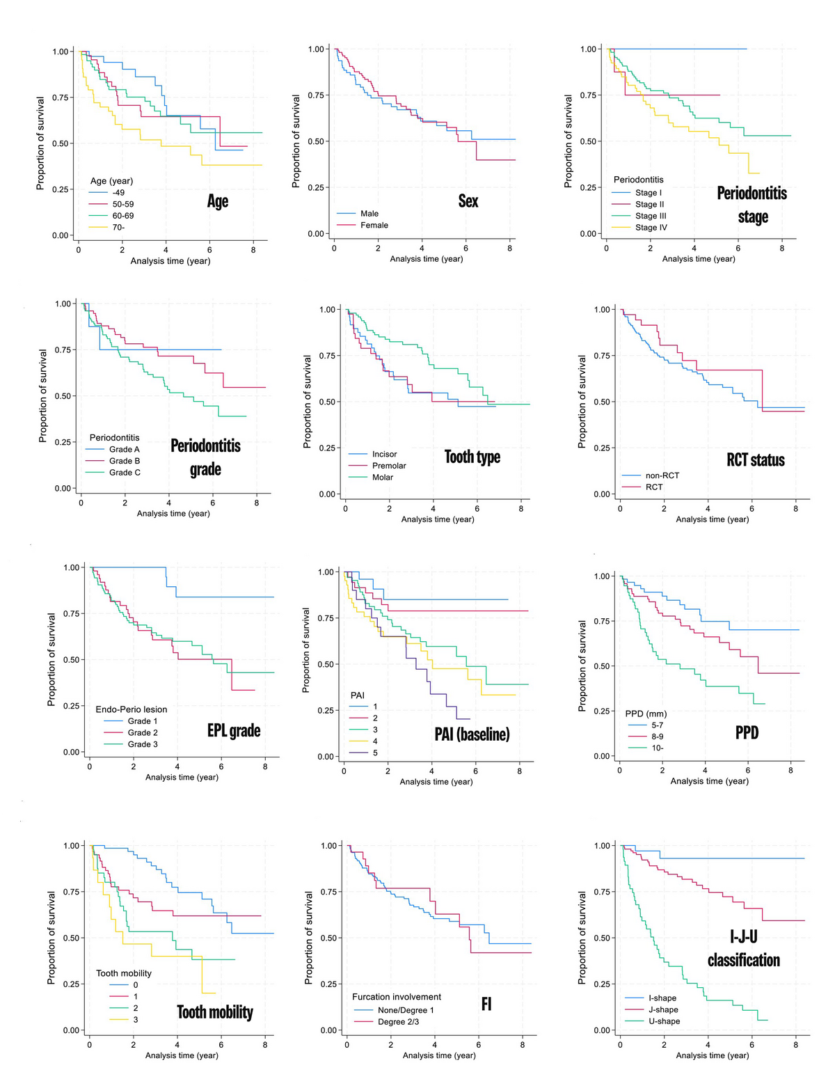
- Tooth survival rate: EPL grade (p = 0.006), PAI (p = 0.001), PD (p < 0.001), tooth mobility (p < 0.001), and I-J-U subclassification (p < 0.001) were significantly associated with the tooth survival rate in the stratified Kaplan-Meier analysis.
Radiographic bone loss morphology:
- The estimated five-year survival rates for I-, J-, and U-shaped lesions were respectively 93.0%, 72.3%, and 13.4% (p < 0.001).
- J-shape (HR 4.44; 95% CI: 1.02–19.31; p = 0.047) and U-shape (HR, 28.06; 95% CI: 6.03–130.51; p < 0.001) were associated with a significantly higher risk of extraction compared with I-shape in the multivariate Cox regression model including the I–J–U classification.
- The model including the I–J–U subclassification showed a better fit (AIC 586.5 vs. 617.9) and a higher five-year predictive accuracy (AUC 0.85 vs. 0.77) compared with the model without it.
Limitations
- The fact that 94.2% of patients suffered from severe periodontitis (stage III or IV) may limit the ability to generalize the findings in relation to patients with less severe periodontitis.
- Only teeth that underwent treatment were included. Teeth extracted without treatment were excluded, which may interfere with results (selection bias, overestimating survival).
- The retrospective design and reliance on two-dimensional radiographs (although the parallel technique was used) may limit diagnostic accuracy compared with three-dimensional imaging (CBCT).
- The prognostic analysis did not include post-treatment changes in clinical and radiographic parameters or adherence to supportive periodontal care (SPC).
Conclusions and impact
- This retrospective study found that age, EPL grade, probing depth, and the radiographic pattern of bone loss served as significant pretreatment predictors of tooth survival in teeth with EPLs.
- The newly proposed I-J-U radiographic subclassification was associated with long-term tooth survival, with U-shaped lesions demonstrating a markedly poorer prognosis than I- or J-shaped lesions.
- The initially high one-year tooth survival rate (83.8%) declined to 58.9% at five years, highlighting the continuous risk of recurrent periodontal breakdown.
These findings support the use of radiographic bone-loss morphology as an adjunct to clinical parameters for the prognosis of teeth with EPLs.
Rapporteurs: Dimitra Adamouli, Veta Chatziangelaki, Rodopi Emfietzoglou, Chrysa Marasli, and Maria-Myrsini Pouliou, supervized by Prof. Phoebus Madianos and Prof. Spyridon Vasilopoulos
Affiliation: Postgraduate programme in periodontology, National and Kapodistrian University, Athens, Greece
With kind permission from Wiley Online Library. Copyright © 1999-2026 John Wiley & Sons, Inc. All rights reserved
EFP partners









Sign up to our newsletter
The EFP publishes a free monthly email newsletter with the latest news about the federation's activities, its publications, and its campaigns.
