
Issue No. 150
Summarized from Journal of Clinical Periodontology, Volume 53, Issue 6, June 2026, 933-946
Editor: James Deschner, chair, EFP scientific affairs committee
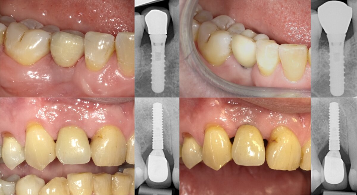
Surgical treatment of peri-implantitis: reconstructive versus open-flap debridement
Authors: Jarno Hakkers, Yvonne C. M. de Waal, Barzi Gareb, Henny J. A. Meijer, Gerry M. Raghoebar
Background
Peri-implantitis is a plaque-associated pathological condition characterized by progressive peri-implant bone loss, increased probing depths, and bleeding and/or suppuration on probing. Although several surgical and non-surgical treatment approaches have been proposed, it remains a challenge to achieve stable, long-term disease resolution.
Surgical therapy is frequently indicated in severe peri-implantitis cases, particularly when deep intrabony lesions are present. Among the available surgical strategies, reconstructive procedures have been introduced to promote bone fill of the defect and improve peri-implant tissue architecture. Various grafting materials and membranes have been proposed to support peri-implant bone regeneration and potentially enhance clinical outcomes.
However, current evidence regarding the superiority of reconstructive approaches over open-flap debridement remains inconclusive. While some studies have reported improved radiographic bone fill and soft-tissue stability following reconstructive therapy, others have failed to demonstrate substantial improvements in inflammatory parameters or disease resolution.
Therefore, further randomized controlled clinical studies are needed to clarify the potential benefits and limitations of reconstructive peri-implantitis surgery.
Aim
The aim of this randomized controlled clinical trial was to evaluate the effect of adjunctive reconstructive therapy, performed during surgical peri-implantitis treatment of three- and four-wall intrabony defects, on clinical and radiographic outcomes at one-year follow-up.
Materials and methods
- A single-blind randomized controlled clinical trial with a one-year follow-up.
- Fifty-two patients presenting peri-implantitis associated with three- or four-wall intrabony defects were randomly allocated to:
- reconstructive surgical therapy (test group; n=27), or
- open-flap debridement (control group; n=25)
- Peri-implantitis was diagnosed according to the 2017 World Workshop criteria and defined by:
- bleeding and/or suppuration on probing, increased probing depths compared with previous examinations, and radiographic bone loss beyond physiologic remodeling.
- In the absence of prior records, peri-implantitis was diagnosed based on probing depths ≥6mm and bone loss ≥3mm apical to the most coronal intraosseous part of the implant.
- Before surgery, all patients received non-surgical peri-implant therapy, including oral-hygiene instructions and mechanical debridement.
- During surgery, full-thickness flaps were elevated and implant surfaces were decontaminated using air-polishing and phosphoric acid.
- In the reconstructive group, peri-implant defects were filled using a mixture of autogenous bone and deproteinized bovine bone mineral covered with a collagen membrane.
- In the control group, open-flap debridement without reconstructive procedures was performed.
- Primary outcomes included peri-implant probing pocket depth (PPD) and radiographic marginal bone level (MBL).
- Secondary outcomes included bleeding on probing (BoP), suppuration on probing (SoP), disease resolution (residual probing depths ≤5mm; BoP in ≤1 probing site; no suppuration on probing), mid-buccal recession, and width of keratinized mucosa.
- Clinical and radiographic parameters were assessed at baseline and during follow-up visits up to 12 months.
- Data were analysed using multilevel mixed-effects models according to the intention-to-treat principle.
Results
- Both treatment approaches resulted in substantial reductions in peri-implant probing depths throughout the 12-month follow-up period.
- Reconstructive therapy was associated with significantly improved radiographic marginal bone levels compared with open-flap debridement at all follow-up visits:
- three months: −0.85 mm (p=0.04)
- six months: −1.12 mm (p=0.01)
- nine months: −1.39 mm (p<0.001)
- 12 months: −1.65 mm (p<0.001)
- The reconstructive group also demonstrated significantly lower mid-buccal recession at all follow-up visits compared with the control group.
- No significant differences between groups were observed for: PPD, BoP, SoP, keratinized mucosa width, or overall disease resolution.
- At 12 months, disease resolution was achieved in:
- 43.8% of patients in the reconstructive group, and
- 44.4% of patients in the control group.
- At the implant level, 19.2% of implants in the reconstructive group and 26.1% in the control group still presented residual probing depths >5mm at 12 months.
- Both treatment approaches resulted in marked reductions in suppuration and plaque accumulation compared with baseline values.
- Subgroup analyses suggested that reconstructive therapy provided greater radiographic benefits in both three- and four-walled defects, particularly regarding the maintenance of the marginal bone level.
- No membrane or graft-related complications were observed in the reconstructive-surgery group.
- Patients treated with reconstructive surgery reported higher frequencies of post-operative pain, mouth dryness, metallic taste, and headache compared with the control group.
Limitations
• The follow-up period was limited to 12 months, which may not be sufficient to evaluate the long-term stability of peri-implantitis treatment outcomes.
• The radiographic bone fill observed in the reconstructive group does not necessarily confirm true re-osseointegration or biologic regeneration.
• The study included different implant systems, prosthetic reconstructions, and previously augmented sites, potentially introducing clinical heterogeneity.
• The primary outcome was modified after the initiation of the study, with greater emphasis placed on probing depth and marginal bone level outcomes.
• Exploratory subgroup analyses according to defect configuration may have been underpowered.
• The implant surface decontamination protocol included air-polishing and the application of phosphoric acid, which may limit reproducibility and comparability with other contemporary treatment protocols.
• Formal patient-reported outcome measures regarding oral-health-related quality of life and aesthetic perception were not assessed.
Conclusions and impact
- Surgical treatment of peri-implantitis resulted in significant clinical and radiographic improvements after one year of follow-up.
- Reconstructive therapy was associated with superior radiographic marginal bone levels and less mid-buccal soft-tissue recession than open-flap debridement alone.
- Despite these radiographic and soft-tissue benefits, reconstructive therapy did not significantly improve inflammatory parameters or overall disease resolution.
- Maintaining complete peri-implant health remained difficult in both treatment groups, highlighting the complexity of peri-implantitis management.
Reconstructive peri-implantitis surgery may be especially beneficial for deep intrabony defects when preserving peri-implant tissue architecture is a treatment objective.
Long-term studies are still needed to determine whether the observed radiographic improvements translate into higher implant survival and long-term peri-implant stability.
Rapporteurs: Tommaso Conforti and Julia Ríos-Barbero, supervised by Professor Mariano Sanz Alonso
Affiliation: Postgraduate programme in periodontology, Complutense University of Madrid, Spain
With kind permission from Wiley Online Library. Copyright © 1999-2026 John Wiley & Sons, Inc. All rights reserved
EFP partners


Sign up to our newsletter
The EFP publishes a free monthly email newsletter with the latest news about the federation's activities, its publications, and its campaigns.