
Classification & Guidelines, Clinical & Translational Research, Article
What needs to be considered in treating gingival diseases and conditions in systemically healthy children and adolescents
18 September 2026
The consensus report of the focused workshop organized by the EFP and the European Academy of Paediatric Dentistry (EAPD) on gingival and periodontal diseases in children and adolescents was published in the July 2026 issue of the federation’s Journal of Clinical Periodontology. At the workshop, three working groups considered systematic reviews and developed their consensus view. Mariano Sanz, who chaired the working group on gingival diseases and conditions in healthy children and adolescents, explains the key findings.
At the focused workshop, the term “periodontal disease” was deemed to include not only conditions limited to the gingival tissues but also those affecting the periodontal attachment apparatus. In its broadest sense, this includes conditions that were or were not induced by dental biofilm in both systemically healthy patients and in those with underlying systemic diseases or conditions that impacted upon the periodontium. It was recognized that the periodontal tissues of children—whether in the primary, mixed, or early permanent dentition—significantly differ anatomically from those of adults (Bimstein et al., 2001; Bimstein and Matsson, 1999).
Periodontal diseases in children and adolescents are common. However, the use of multiple measures to examine and record disease, together with the historical lack of consistency in case definitions, hinder the interpretation and comparison of prevalence and incidence estimates for periodontitis found in the literature. The workshop’s working group 2, chaired by Mariano Sanz and Phoebus Madianos, focused on gingivitis and gingival diseases, and the systematic review compiling the evidence was led by Georgios Tsilingaridis and Rodrigo López (Tsilingaridis et al., 2026).
Although dental-biofilm-induced gingivitis is the most common manifestation of gingival diseases in systemically healthy children and adolescents, gingival diseases may exhibit several distinctive features that differ from those observed in adults, and the presence of local and systemic determinants strongly influences these conditions.
Dental-plaque biofilm, which contains microorganisms that cause gingival inflammation, is the main local cause of inflammation in children and adolescents. This condition is frequently associated with inadequate oral-hygiene practices. In this population, the presence of local factors has been linked to increased plaque accumulation, which significantly contributes to gingivitis in children and adolescents. Among these local factors, plaque-retentive dental-plaque-biofilm factors include:
(a) orthodontic appliances
(b) open dental caries lesions
(c) overhanging restorations
(d) anatomical issues
(i) crowded and misaligned teeth
(ii) partially erupted teeth
(iii) food impaction
(iv) dental calculus.
Similarly, the presence of mouth breathing—which frequently occurs in children with allergies or adenoid hypertrophy—leads to the drying of the gingival tissues, reduced plaque removal by saliva, and heightened inflammation.
What is the prevalence?
Gingivitis induced by dental biofilm is common in children and adolescents, with its incidence increasing with age. In epidemiological studies, the prevalence data for gingivitis is challenging to interpret because of variations in studies from different countries. The overall estimate is about 50%, but published results are highly heterogeneous, and statistics derived from the community periodontal index (CPI) in two studies vary from 18% to 92%. (Tsilingaridis et al., 2026).
Clinically, gingivitis is characterized by redness, swelling, and bleeding of the gums; it is generally painless and without loss of alveolar bone. Gingival inflammation is typically less frequent and less severe in younger children than in older ones, possibly because of the greater thickness and height of the gingival tissues. However, depending on the presence of specific local or systemic determinants, various gingival conditions have been identified, although their pathophysiology is similarly the consequence of the accumulation of dental plaque. For example, during the eruption of primary or permanent teeth, there is usually localized inflammation around erupting teeth. Similarly, hormonal changes, usually around puberty, influence the response to plaque exposure, leading to a higher prevalence and severity of gingivitis, typically presenting with significant enlargement of the interdental papillae.
Diagnosis is based on visual examination of gingival inflammation, including evaluation of changes in the colour, contour, consistency, and texture of the gingival margins and papillae; detection of bleeding on brushing; and periodontal probing. However, periodontal probing should not be used in children, because of the immaturity of their periodontal tissues, or in children with mixed dentition including erupting teeth. These methods need to be adapted to each child’s developmental stage and level of co-operation. Radiographic examinations should be undertaken only when diagnosing caries lesions or when there is a clear clinical indication.
While the 2018 classification of periodontal diseases and conditions established a threshold of 10% bleeding on probing (BOP) sites for defining gingivitis in adults, there is a consensus that this threshold is not suitable for defining gingivitis in children and adolescents until there is a clear understanding of the prevalence and distribution of gingivitis in this population.
Systemic modifying factors often interact with local factors to enhance plaque accumulation and/or response to plaque accumulation. The most relevant factors in this population are the hormonal changes during puberty and being overweight or obese—both associated with increased low-grade inflammation—or an increased gingival response to plaque. Environmental factors—such as exposure to tobacco smoking or the intake of carbohydrate-rich diets—are less common in children, and the evidence is very scarce.
Focus of treatment
The treatment of dental-biofilm-induced gingivitis in children and adolescents focuses on controlling dental-biofilm buildup and reducing gingival inflammation. In most cases, improved oral hygiene and professional mechanical plaque removal (PMPR) lead to a positive response. When inflammation persists, underlying systemic conditions should be assessed. Depending on age, oral hygiene should be supervised by parents or caregivers.
The main recommendation is to brush twice daily with either a manual or powered toothbrush using a fluoride toothpaste. One should consider using agents to visualize plaque to increase motivation and facilitate more effective removal. Interdental cleaning methods should be tailored to age. The use of antimicrobial toothpastes or mouthrinses should be age-appropriate and recommended only when gingival inflammation does not revert with mechanical oral-hygiene methods, which would be the case mainly in children and adolescents using orthodontic appliances. After treatment, it is important to re-evaluate gingival health and adjust the treatment plan as necessary, mainly through reinforcing oral-hygiene instructions.
Orthodontic treatment can sometimes lead not only to dental-biofilm-induced gingivitis but also to gingival enlargement and gingival recession. The primary preventive strategies during orthodontic therapy involve coaching in oral hygiene, including detailed instructions for proper brushing techniques around brackets and wires, and using special orthodontic brushes, interdental brushes, and floss threaders. The primary professional intervention is PMPR. More frequent dental visits are recommended to evaluate gingival health and should always be tailored to the patient's needs.
When gingival hyperplasia or enlargement occurs, its cause is often attributed to plaque accumulation around orthodontic appliances. Therefore, its management focuses on improving oral hygiene and PMPR. In severe cases, the orthodontist should be consulted to discuss the possibility of halting orthodontic therapy and removing the appliances to reinforce oral-hygiene practices. Depending on the specific situation, additional surgical-tissue excision may be necessary, either during or after orthodontic treatment. Drug-induced gingival enlargement in children and adolescents may also be associated with the use of certain medications (anticonvulsants/antiepileptics, immunosuppressants, and calcium channel blockers). Its management typically involves medication adjustments in consultation with the prescribing physician, enhanced oral hygiene, professional dental care and—in severe cases—surgical intervention.
Acute infections and reactive processes
Acute infections in children and adolescents can be very distressing, especially for young children, because of the pain and discomfort that often lead to decreased fluid intake and an increased risk of dehydration. Treatment focuses primarily on supportive care, including pain management, maintaining hydration, and preventing secondary infections. In severe cases, hospitalization for parenteral fluid administration should be considered. The most common acute gingival infections affecting children and adolescents are viral infections, the most frequent of which are: herpetic gingivostomatitis; hand, foot, and mouth disease (HFM) caused by Coxsackievirus A16; herpangina; and varicella zoster.
Reactive processes, although rare in children, can manifest as localized, non-neoplastic growths that affect the gingival tissues (fibrous epulides), typically associated with chronic irritation or trauma. Additionally, a pyogenic granuloma (a vascular epulis) can occur during puberty. Management usually involves surgical excision with histopathological examination to confirm the diagnosis. Removing potential irritants is crucial to prevent recurrence.
Traumatic lesions of the gingival tissues most often result from mechanical trauma: abrasion or laceration from tooth brushing or self-inflicted by, for example, pencil-biting. These lesions typically present with pain, bleeding, and inflammation, and sometimes with ulcerations, erosions, gingival clefts, and gingival recession. Treatment focuses on eliminating the causative factor(s), supporting tissue healing, and preventing recurrence through patient education and adjusted oral-hygiene techniques.
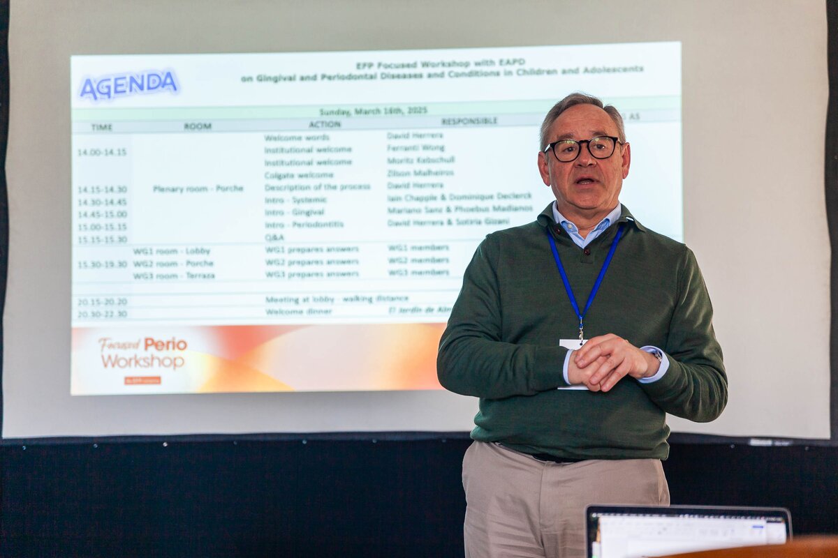
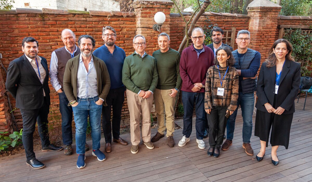
The EFP/EAPD focused workshop on gingival and periodontal diseases in children and adolescents was held in Madrid, Spain, in March 2025. The workshop was sponsored by Colgate-Palmolive, one of the EFP’s partners. The consensus report was published in the July 2026 edition of the Journal for Clinical Periodontology.
Bibliography
Bimstein E et al., 2011.Periodontal and gingival health and diseases: children, adolescents, and young adults (London: Martin Dunitz).
Bimstein E and Matsson L. 1999. Growth and development considerations in the diagnosis of gingivitis and periodontitis in children. Paediatric Dentistry 21(3): 186-91
Chapple I, Gizani S, Sanz M, et al. 2026. “Gingival and Periodontal Diseases and Conditions in children and Adolescents: Consensus Report.” Journal of Clinical Periodontology 53(7): 1068-1099. doi: 10.1111/jcpe.70033. Epub 2026 Apr 15.
Chapple I, Gizani S, Sanz M, et al. 2026. “Gingival and Periodontal Diseases and Conditions in children and Adolescents: Consensus Report.” European Archives of Paediatric Dentistry 28. https:// doi. org/10. 1007/ s4036 8-025-01154-y.
Tsilingaridis, G, Tewari N, Seremidi K, Papaioannou W, López R. 2026. “Dental Biofilm–Induced Gingivitis in Children and Adolescents Without Known Systemic Involvement: A Systematic Review.” Journal of Clinical Periodontology 53(7): 1226–1252.
Biography
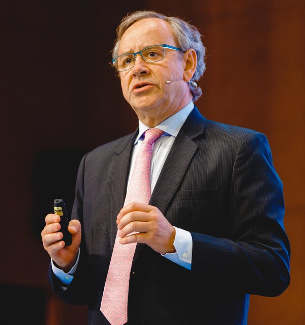
Mariano Sanz is the director of postgraduate periodontology (EFP-accredited programme) and the ETEP (Etiology and Therapy of Periodontal and Peri-Implant Diseases) research group at the Complutense University of Madrid (UCM) in Spain, and a Type II professor at the University of Oslo in Norway. Professor Sanz has published over 545 scientific publications (ORCID # 0000-0002-6293-5755; h-index 89), and is an international lecturer on topics in periodontology, implant dentistry, and dental education. He has served the EFP as president, secretary general, and chair of the workshop committee. He has received six honorary doctorate degrees from the University of Goteborg (Sweden), Coimbra (Portugal), San Sebastián (Santiago de Chile), the University of Buenos Aires (Argentina), the Medical University of Warsaw (Poland), and the University of Athens (Greece).
EFP partners









Sign up to our newsletter
The EFP publishes a free monthly email newsletter with the latest news about the federation's activities, its publications, and its campaigns.
